Learn From the Dental Industry's TOP LEADERS!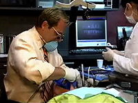
Sit Chairside withDr. Dennis WellsCreator ofDURAthin® Prepless Veneers- OR -
|
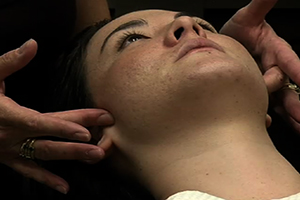
A Joint ExamPart 4 - Doppler auscultation
Instructors:
Dr. Brady describes why ausculatation of the joint is an important part of joint assessment and then demonstrates the use of a doppler.
So a last piece of this learning opportunity about the joint exam and joint diagnosis is being able to auscultate the joints. So we talk about palpable joint sounds. And I wasn't aware of anything palpable in Katie's condition but we want to listen. Because there are lots of joint sounds that happen that I will not be able to detect under my finger and I might not get from Katie in history. So how are we going to auscultate. We're going to do that with a Doppler. You can also do this with a stethoscope although I will tell you that it's much harder to hear with a stethoscope and one of the things I love about a Doppler is that the patient gets to hear exactly what I'm hearing as we do the exam. It's the same technology as OB/GYN used to hear on unborn baby's heartbeat. It just amplifies sounds. And I tell the patient that and most patients are pretty familiar with that technology. It's pretty every day. So Katie would it be okay if I listen to this right joint with a Doppler? Yes. All right. So let's just start by talking about the Doppler. And so lots of places to get a Doppler. This happens to be the one I used in my office that I got from Great Lakes Orthodontics. One of the things that I want to do first with a Doppler is I want to make sure and test the batteries. Dopplers can very easily create a lot of false positive background noise as the batteries are starting to die. So the first thing that I do is simply turn the Doppler on and listen. And what you'll notice is you can definitely hear some white noise, some background noise but we don't hear anything from crunchy or staticky. If we heard that without teaching anything what it basically means is its time to replace the 9 volt battery in this. So that's the first thing. The next thing I'm going to do is I'm going to explain to Katie exactly what I'm going to hear. So Katie a normal healthy joint is going to be totally quiet under a Doppler. All you'll hear is this is background noise. If we hear anything other than that background noise then you and I will want to talk about what that means, what's going on inside your joints. Okay. We'll actually going to put some jelly on this just like you do when you to hear a baby's heartbeat and sometimes it's a little bit cold and it will definitely be a little bit messy. I'll try to get your hair out of the way as much as possible. So now she knows what we're going to do and I want to try to isolate some different things when I do this. We talk in the other piece about joint diagnosis how important it is to understand where we hear the joint noise. Is it on rotation? Telling me there's something going on with the medial aspect of the disc or is it on translation. Telling me it's a lateral aspect of the disc. Those are two totally different patients. They have different risk from a restorative perspective and they have different futures and outcomes from the standpoint of treating their joints and getting them asymptomatic. So that's going to be important. How I'm going to do that is I'm going ask Katie to do some very specific movements for me so that I can isolate those sounds. So as we get started what we're going to do is first I'm going to make sure the Doppler is on but I'll turn the noise all the way down so it's not distracting. I actually put the Doppler jelly right on to the head of the Doppler and as you can see you want a really generous amount. Okay. Really critical that the actual head of the Doppler never touch Katie's skin, that the sound is being communicated through the jelly so I need at least a good quarter inch of that between her skin and the head of the Doppler. Next thing I'm going to do is I'm going to have her turn her ahead away from me just a little bit and I am going to just sort of sneak her hair out of the way as best as possible. I want to remind myself where the lateral pole of her condyle was. So I'm going to actually ask her to open and close once and close and find the lateral pole of her condyle. Now I'm going to actually put the Doppler in place just behind that and as you can see not touching her skin. [And now I can hear her superficial temporal artery that tells me I'm in the right place. So now what I'm going to do is I'm going to tip the Doppler forward until I lose the sound of the artery and I'm going to actually ask Katie to open and close three times so I hear a lot of scratching so I that I can get to the loudest spot.] Perfect. So now I'm going to turn the sound down for a second so we can talk. What you'll notice is I ask Katie to move quickly because when she moves quickly I hear a lot of crunching noises. That's actually turbulence in the synovial fluid because she's moving so quickly. When I actually do the Doppler exam if she moves too fast I will think I hear crepitus but it's a false positive. So I'm going to have actually control her movements now. So the first thing I want to do is listen to rotation. So I'm going to just simply Katie to only open about a quarter of an inch and when she closes not to actually touch her teeth together because you can often think that that's a pop or a click when you hear the clack of the teeth coming into contact. So are you ready Katie? All right. [So just about a quarter of an inch, and close without touching and open and close and open and close. All right. So Katie's joint is totally normal in rotation. Another way to isolate rotation would be to have her do a working side movement so I can could ask Katie to move her jaw toward me. So Katie now go slowly about a second over to the right and back and over to the right and back. One more time and back. Okay. Now I can look at this and do translation. So now what I'm going to do is I'm going to have Katie open to the full extent but I'm only going to pay attention after she gets out of rotation. So Katie take about a second to open and a second to close but open to the full extent that you can. One 1,000 open and close and one 1,000 and close.] So this is a perfect example of one of those unexpected joint sounds. So Katie's history was negative for a pop or a click. We didn't feel a pop or a click but Katie has very definitive crepitus on translation telling us without a pop or a click that she's off the lateral pole of the disc on the side and it doesn't reduce. So we would call this a lateral pole disc displacement without reduction. Now those findings of her load test become even a little more fuzzy because now we do know she is capable of pinching retrodiscal tissue although not classical in a seated position but in excursion. So a finding, her telling me that she has discomfort on this right joint in a lateral movement would not be unexpected. So let's verify translation by having her do a translatory movement on this side meaning moving away from the Doppler. [So Katie I'm going to have you move to the left about a second over and a second back and second over and a second back.] So she has that same crepitus on translation in her lateral movement as she does in her wide opening so then we're just going to remove the Doppler gel. And consequently if we would doing a full exam on Katie we would repeat that on the other side. This for me is, so you all know Katie and I did not stage this, do this ahead of time. But this is a classic example of why I listen to every joint in my practice because Katie is a patient who to the best of her knowledge did not have any problems with the condyle disc assembly. We weren't able to get to the bottom of that to her history and yet listening she and I are both aware of an underlying issue with this joint. What does that mean for us moving forward? Well it tell us there's something going on there that we might need to look at and figure out how do we get rid of the inflammation in this joint. How do we actually work with this so that we can make sure this is a stable condition? As I said in the other video, we don't treat joint noise. Joint noise tells us about historical damage. At some point in the past, Katie lost the lateral pole of the disc on this right joint and we actually known unbeknown to her there was some kind of a whiplash injury so maybe that's what it was. We also know Katie does do a little bit of clenching so potentially its micro trauma. But the crepitus tells us a damage that happens in the past. Now my job with Katie is to figure out is that damage still happening? Is it underway so it could potentially get worst? And if so what could be we do to stabilize the situation or is it currently stable and once it is stable Katie could stay just like this and her joint findings could stay exactly the same with the absence of the positive load test and the inflammation for the rest of her life without concern. So that will be the next thing she and I have to figure out together on the top of the bite appliance. So with this, I want to thank you all for being with us today. My encouragement is to go back to your practice, try some of these things out. You know you have a team of your own and one of the things to do is offer them the opportunity to have a comprehensive exam and sit in a chair so that you can learn, but it also gives them experiences that they can then talk to your patients about. Just a reminder a couple of companion videos that you might want to watch if you've not already with this one. One is the joint and the muscle exam and then of course the joint diagnosis and then anterior bite plate. I talked quite a bit about that will be our next step is to fabricate Katie an anterior bite plate and there is a video showing that fabrication technique. So thank you for being with me and Katie and Crystal and here on HDiQ. |
 FAGD/MAGD Credit Approval does not imply acceptance by a state or provincial board of dentistry or AGD endorsement 7/31/2018 to 7/31/2021 Provider ID# 317928
 Submitting...